
One of the most common challenges when learning DVT ultrasound is knowing exactly how much pressure to apply during vein compression. Applying too little pressure or compressing the vessel at an angle can make a normal vein appear noncompressible and potentially lead to a false-positive finding.
In this Gulfcoast Ultrasound Institute (GCUS) Ultrasound Hot Tip, Karen Avgush, MD, demonstrates a simple technique for recognizing adequate compression: instead of focusing only on the vein, watch the adjacent artery.
How Much Pressure Should You Apply During DVT Ultrasound?
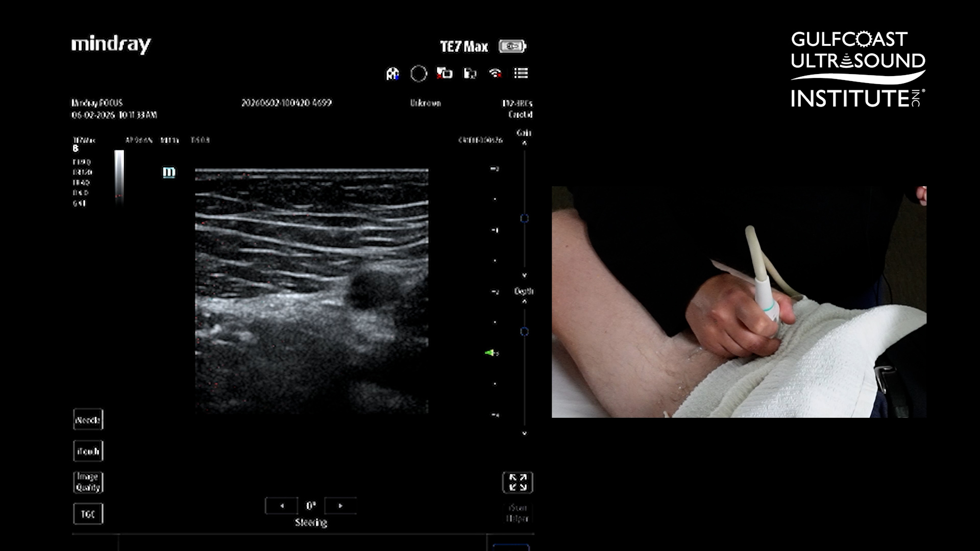
During a compression ultrasound examination, apply steady pressure until you see the adjacent artery begin to compress slightly. This provides a useful visual cue that adequate pressure is being applied.
If the artery begins to deform slightly and the vein completely collapses, that represents a normal compression response. The vein should essentially disappear while the artery remains visible and pulsatile.
Why Should You Watch the Artery During Vein Compression?
The artery can serve as a practical reference point for determining whether enough pressure has been applied. Because the artery has a thicker wall than the adjacent vein, the vein should collapse before significant arterial compression occurs.
A simple way to remember the technique is: artery slightly compressed, vein completely disappears.
What Does a Noncompressible Vein Mean on DVT Ultrasound?
If the adjacent artery begins to compress slightly but the vein remains partially open despite adequate compression, the finding should raise concern for possible DVT. The ultrasound finding should be evaluated as part of the complete examination and clinical picture.
What Can Cause a False-Positive DVT Ultrasound Finding?
Two common technical errors can create the appearance of a noncompressible vessel: not applying enough pressure and compressing at an angle.
If the transducer is not perpendicular to the vessel, pressure may be applied obliquely rather than directly through the vein. This can prevent complete collapse and make a normal vessel appear abnormal.
How Should the Probe Be Positioned for DVT Compression Ultrasound?
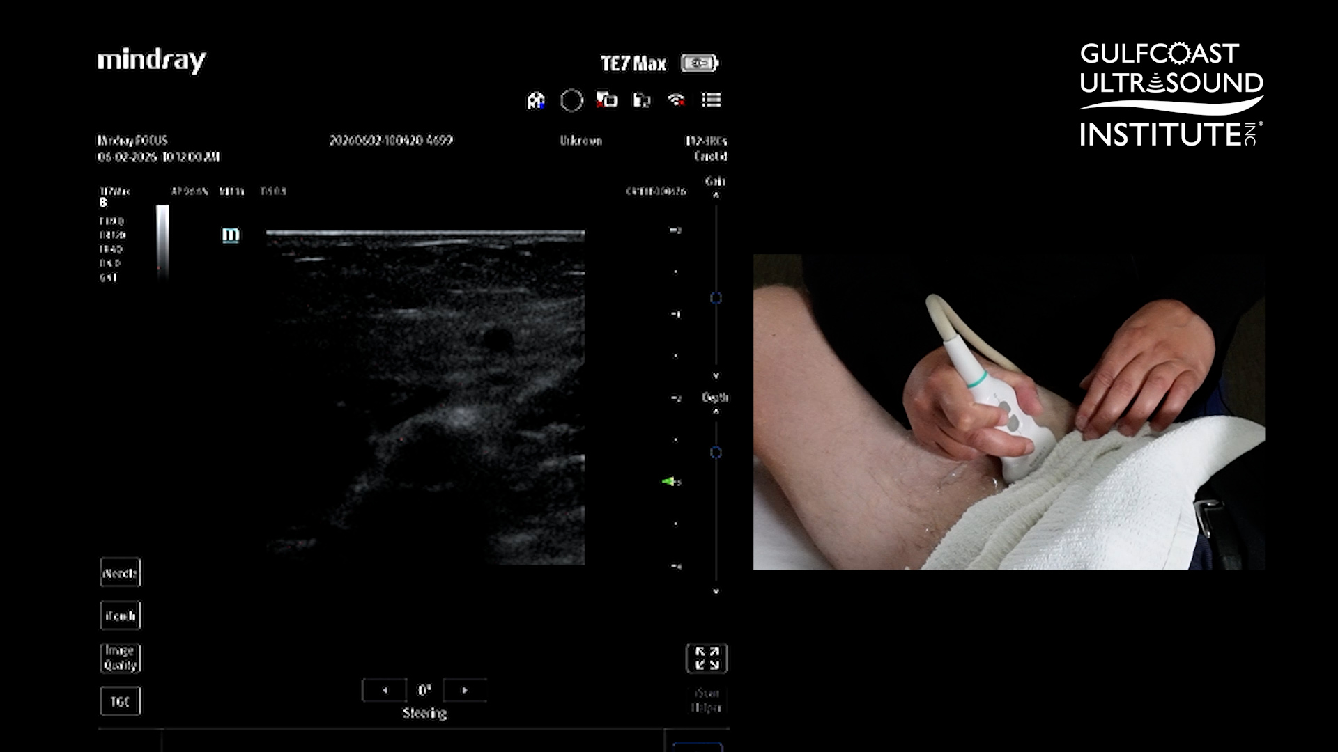
Before compressing, make sure the vein is clearly centered on the ultrasound screen and position the transducer perpendicular to the vessel. Then apply pressure directly downward while watching both the vein and the adjacent artery.
Quick Guide: DVT Ultrasound Compression Technique
- Center the vein and adjacent artery on the ultrasound image.
- Keep the transducer perpendicular to the vessel.
- Apply steady downward pressure.
- Watch the adjacent artery as you compress.
- Continue until the artery begins to compress slightly.
- Confirm that the vein completely collapses.
- If the vein remains partially open despite adequate compression, evaluate for possible DVT.
Using the same systematic technique at each compression point can improve consistency and help reduce errors caused by insufficient pressure or improper transducer positioning.
Why Is Proper DVT Compression Technique Important?
Compression is a fundamental component of ultrasound evaluation for lower-extremity DVT. Accurate technique is essential because incomplete compression can be an important sonographic finding, but technical factors can also mimic noncompressibility.
Learning to recognize adequate pressure, maintain a perpendicular probe position, and keep the vessel centered can help clinicians perform more reliable compression examinations.
Learn DVT Ultrasound Techniques in Person at Gulfcoast Ultrasound Institute
Want to take this Hot Tip from the screen to the scan lab?
Techniques like DVT compression ultrasound can be learned and practiced during Gulfcoast Ultrasound Institute live ultrasound courses and hands-on scanning workshops. GCUS combines comprehensive ultrasound education with extensive hands-on scanning under the guidance of experienced ultrasound faculty.
Hands-on scanning sessions feature an industry-leading 3:1 or less participant-to-instructor ratio, giving participants opportunities to practice image acquisition, probe positioning, scanning protocols, image optimization, DVT evaluation, and other point-of-care ultrasound applications with direct faculty guidance.
Training options are available in multiple educational formats, including live courses, blended ultrasound courses, online education, and hands-on scanning workshops.
Ready to strengthen your DVT and point-of-care ultrasound scanning skills? Explore Gulfcoast Ultrasound Institute ultrasound courses and find the training option that fits your clinical practice.