Ultrasound-guided nerve blocks have become a staple in regional anesthesia and pain management. When performed correctly, they're efficient, targeted, and greatly improve patient comfort and outcomes. Today, we’re walking through a practical and approachable technique for scanning the peripheral nerves of the upper extremity. Whether you're a beginner or brushing up on your skills, this step-by-step breakdown will help you navigate the anatomy and get confident with your probe placement.
Start with the Ulnar Artery as Your Landmark
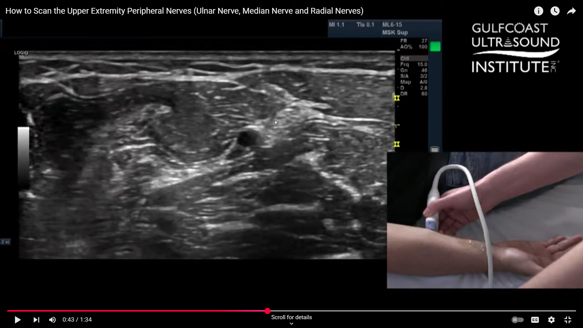
The ulnar artery is your gateway. When scanning the forearm, begin by locating this structure. It’s typically easy to spot and sits right in the center of your screen when you're in the correct transverse view. Once you've found the ulnar artery, the ulnar nerve will be close by, running right alongside it, particularly at the distal end near the wrist. At this level, they’re nearly touching.
Scan Proximally to Watch the Nerve Diverge
As you slowly scan more proximally, up toward the elbow, you’ll notice the ulnar nerve begins to pull away from the artery. Around two-thirds of the way up the forearm, the nerve becomes more distinguishable and easier to isolate. This separation is helpful when prepping for a nerve block, as it creates a clearer visual window for needle guidance.
Identify the Median Nerve from the Same View
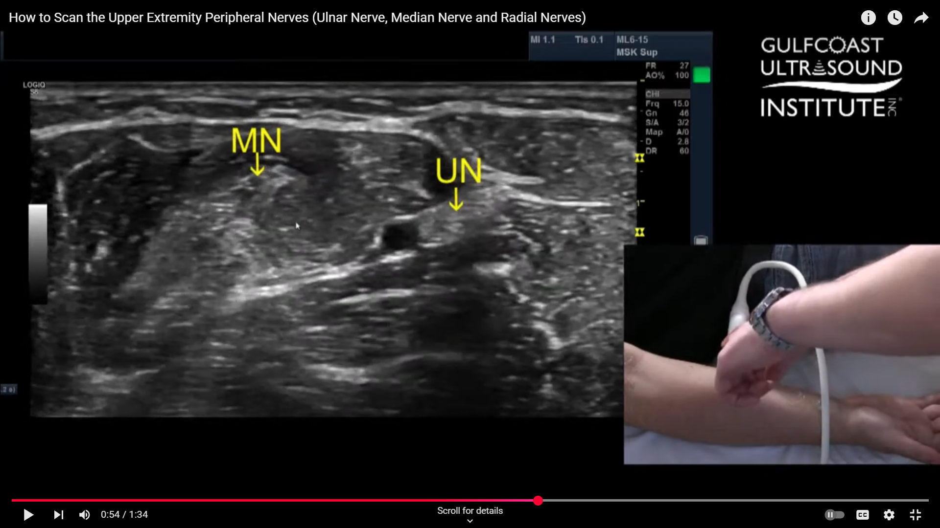
Once you're in that same mid-forearm area, glance medially. You’ll often spot the median nerve in that same imaging plane. With a bit of finesse, you can line up your probe to visualize both the ulnar and median nerves simultaneously. This is a huge advantage, because with one well-placed injection, you can block both nerves at once.
Locating and Blocking the Radial Nerve
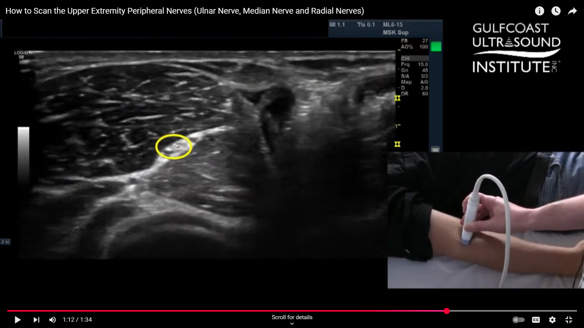
Now for the radial nerve. This one behaves a bit differently. It splits just distal to the elbow, so you'll want your patient to slightly rotate their forearm into a "thumbs-up" position. This hand orientation gives you better access and a cleaner acoustic window. Place your probe along the mid-arm and start sliding it laterally. As you angle and tilt your probe slightly, you’ll see the radial nerve pop up between two muscle layers, nestled right in the fascial plane. It’s a distinct and satisfying view once you catch it.
The Complete Upper Extremity Block
When you’ve successfully identified and blocked the radial, ulnar, and median nerves, you’ve essentially achieved a complete hand block. This comprehensive approach can be extremely beneficial for patients undergoing procedures on the hand or wrist. The ability to manage pain effectively while minimizing systemic medication is a valuable tool for any clinician.
Final Thoughts
That’s your hot tip of the day from Gulfcoast Ultrasound Institute. These foundational skills are key for anyone performing upper extremity nerve blocks. The better you get at visualizing nerves in real time, the more confident and effective you’ll become. Now, it’s your turn to practice and master the technique.
Ready to Build Your Ultrasound Skills?
If you're ready to take your ultrasound training to the next level, we’re here to help. Call the Gulfcoast Ultrasound Institute at 727-363-4500 for all of your ultrasound education needs. We’re conveniently located at 111 2nd Ave NE, #800 St. Petersburg, FL 33701, and we offer a wide range of in-person and online training options to fit your schedule and career goals.
Whether you’re looking to refine your nerve block technique or expand into other specialties, you’ll find expert instruction and real-world guidance at Gulfcoast Ultrasound. Reach out today, we look forward to helping you grow your skillset and confidence.