Proper Color Doppler and Pulsed-Wave Doppler optimization is essential for obtaining diagnostic-quality ultrasound images, accurate blood-flow information, and reliable velocity measurements.
In this Gulfcoast Ultrasound Institute (GCUS) Ultrasound Hot Tip, Emily Downs demonstrates how to optimize Color Doppler and Pulsed-Wave (PW) Doppler ultrasound, using the carotid artery as an example. The same systematic principles including optimizing the 2D image, adjusting the color box and PRF, positioning the sample volume, and correcting the Doppler angle are fundamental skills that can be applied across many diagnostic and point-of-care ultrasound examinations.
What Is the Best Way to Optimize Doppler Ultrasound?
Doppler ultrasound should be optimized systematically rather than by adjusting individual controls at random.
A practical sequence is:
Optimize the 2D image > Optimize Color Doppler > Position the PW Doppler sample volume > Correct the Doppler angle > Optimize the spectral waveform > Obtain measurements
Starting with a properly optimized 2D image creates the foundation for more accurate Color and Spectral Doppler evaluation.
Step 1: Optimize the 2D Ultrasound Image
Before activating Color or Pulsed-Wave Doppler, optimize the 2D B-mode ultrasound image.
Adjust the Ultrasound Gain
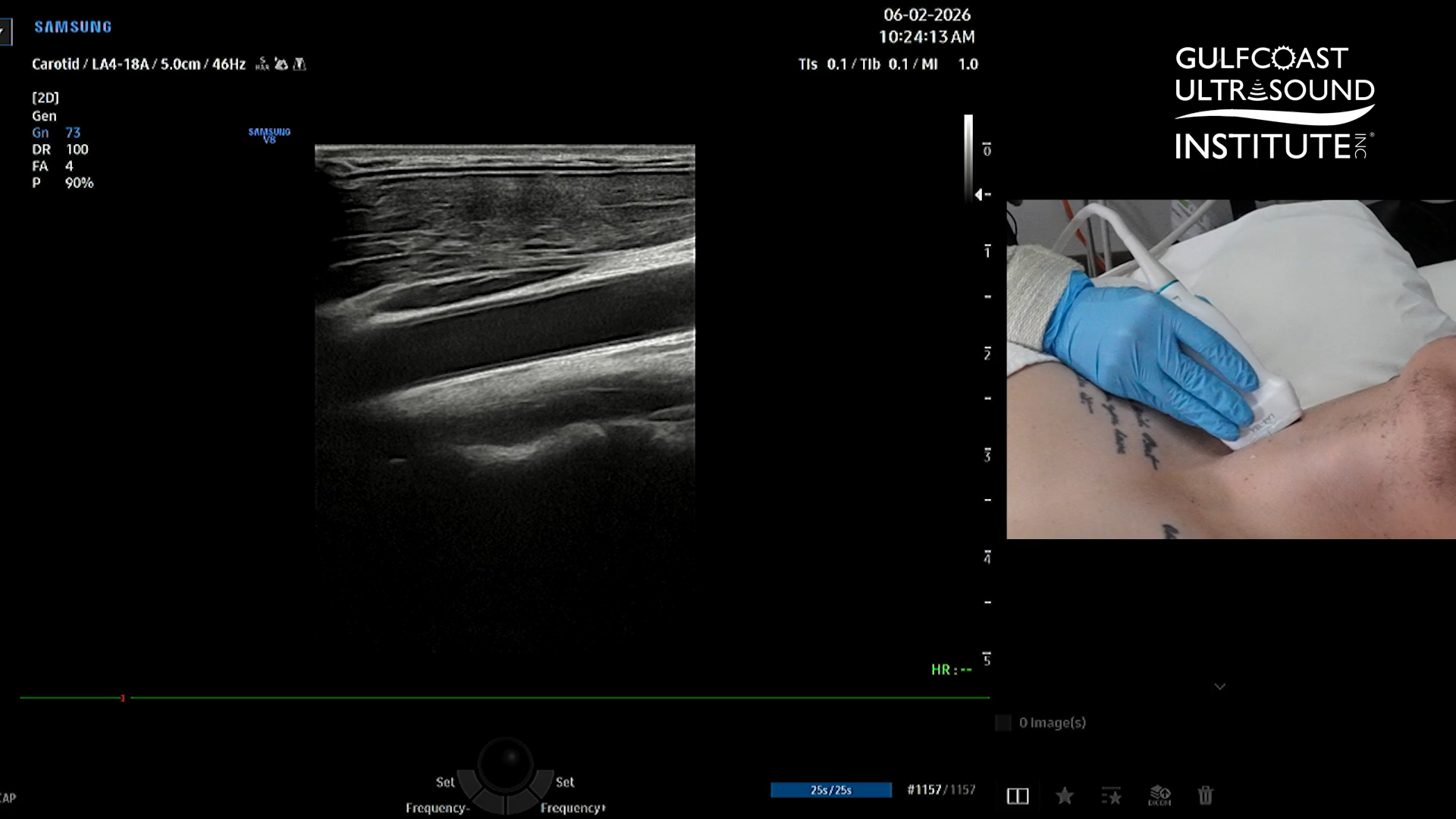
Gain controls the overall brightness of the ultrasound image. Too much gain can introduce unnecessary echoes and reduce image clarity, while insufficient gain may prevent important structures from being adequately visualized.
In this carotid ultrasound example, decreasing the gain produces a cleaner image and improves visualization of the vessel.
Adjust the Imaging Depth
Set the imaging depth so the anatomy of interest is appropriately positioned within the ultrasound display.
For this examination, the carotid artery should be clearly visualized without including unnecessary tissue deep to the vessel. Reducing excessive depth can also improve visualization of the anatomy being evaluated.
Position the Focal Zone
The ultrasound focal zone should generally be positioned at or slightly below the structure of interest.
Correct focal-zone placement helps optimize lateral resolution at the depth where the sonographer needs the greatest image detail.
Step 2: How Do You Optimize Color Doppler?
After obtaining a properly optimized 2D image, activate Color Doppler.
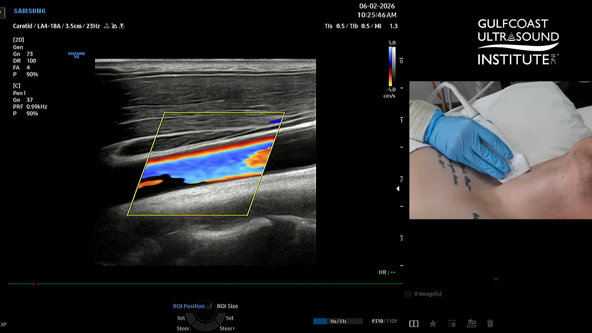
Color Doppler optimization includes appropriately positioning and steering the color box and adjusting settings such as the velocity scale or pulse repetition frequency (PRF).
Steer the Color Doppler Box
The color box should be steered appropriately relative to the direction of blood flow.
When evaluating a vessel such as the carotid artery, steering the color box in the same direction as the vessel will create a more favorable Doppler angle and improve visualization of blood flow.
Adjust the PRF or Velocity Scale
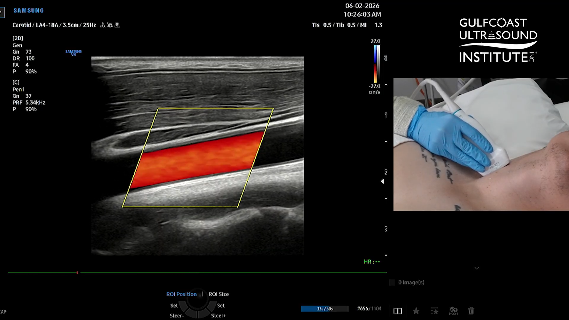
Next, evaluate the color-flow image for aliasing.
Color Doppler aliasing occurs when the Doppler frequency shift exceeds the Nyquist limit. It may appear as an abrupt color change within flowing blood and can potentially be mistaken for a change in flow direction.
Increasing the pulse repetition frequency (PRF) or velocity scale increases the range of velocities that can be displayed and can help eliminate unwanted aliasing.
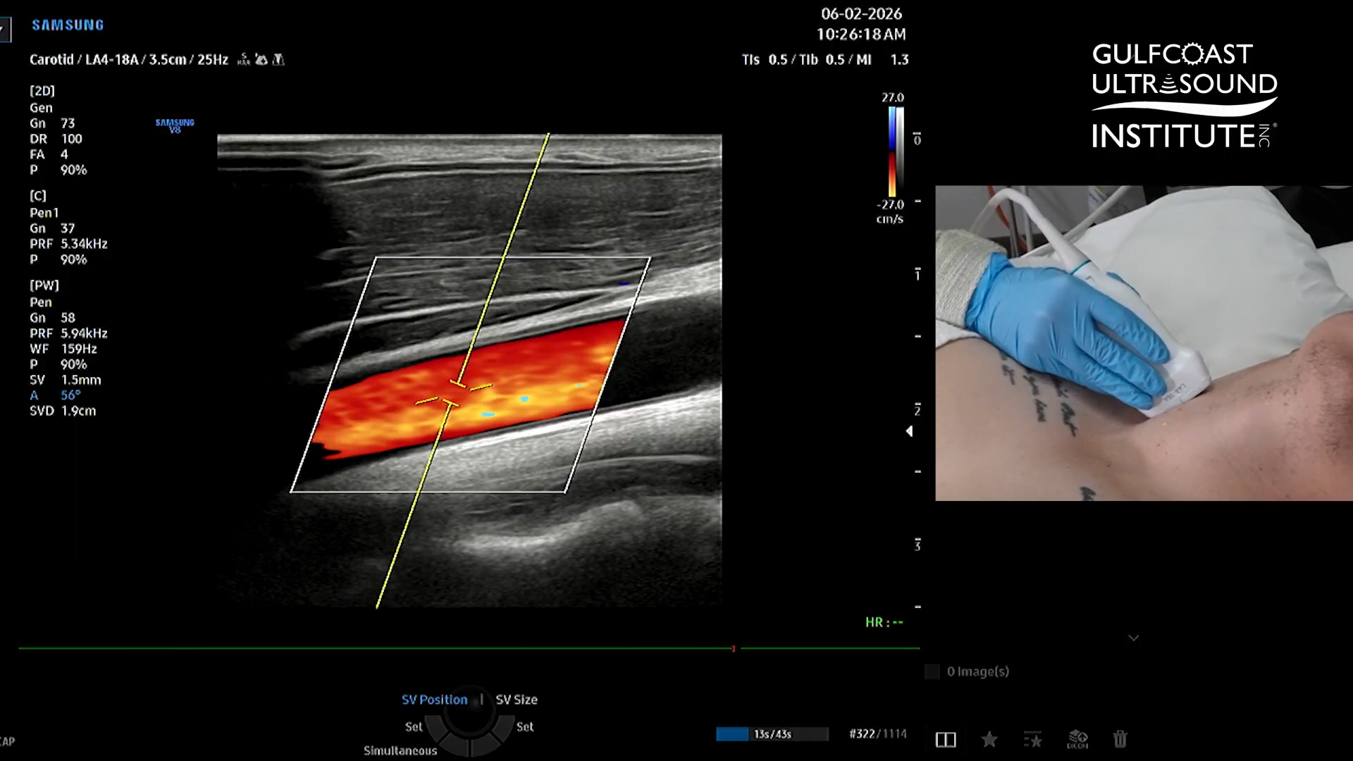
The objective is to select a velocity scale appropriate for the blood-flow velocities being evaluated while maintaining sufficient sensitivity to visualize the flow of interest.
What Is PRF in Doppler Ultrasound?
Pulse repetition frequency (PRF) refers to the number of ultrasound pulses transmitted by the system per second.
In Doppler imaging, PRF plays an important role in determining the range of blood-flow velocities that can be displayed without aliasing.
A PRF that is too low for the velocity being evaluated can result in aliasing. Increasing the PRF or velocity scale allows higher velocities to be displayed appropriately.
PRF must therefore be adjusted according to the expected blood-flow characteristics of the vessel or structure being examined.
Step 3: How Do You Optimize Pulsed-Wave Doppler?
Once Color Doppler has been optimized, activate Pulsed-Wave Doppler, also commonly called PW Doppler or Spectral Doppler.
Place the PW Doppler sample volume within the lumen of the vessel at the location where the velocity measurement will be obtained.
Proper sample-volume placement helps ensure that the spectral waveform represents blood flow from the intended region.
Step 4: Correct the Doppler Angle
Accurate Doppler angle correction is particularly important when calculating blood-flow velocity.
Align the Doppler cursor with the direction of blood flow. In a relatively straight vessel such as the carotid artery, the angle-correction cursor should be positioned parallel to the vessel walls and direction of flow.
Because calculated velocity is dependent on the Doppler angle, improper angle correction can significantly affect the resulting velocity measurement.
Step 5: Optimize the Spectral Doppler Waveform
After activating PW Doppler, evaluate the spectral waveform before taking measurements.
The waveform should be clearly displayed and appropriately sized within the spectral window so the entire tracing can be evaluated.
If the waveform is too large or extends beyond the display, increasing the PRF or velocity scale can reduce its displayed size and allow the complete waveform to be visualized.
An appropriately optimized waveform makes interpretation easier and supports more accurate placement of measurement calipers.
How Is Peak Systolic Velocity Measured with Doppler Ultrasound?
Once the spectral Doppler waveform is properly optimized, measurements such as peak systolic velocity (PSV) can be obtained.
Peak systolic velocity represents the maximum blood-flow velocity occurring during systole within the sampled vessel.
In Emily’s carotid artery demonstration, the optimized PW Doppler waveform produces a peak systolic velocity measurement of approximately 120 cm/s.
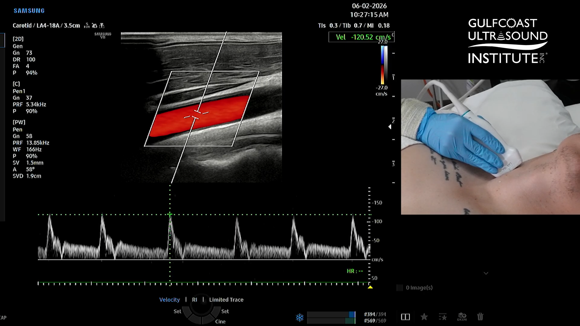
Importantly, measurements should be performed after the Doppler image and waveform have been properly optimized. Accurate measurements depend on appropriate image acquisition, sample-volume placement, angle correction, and Doppler settings.
Quick Guide: Color and Pulsed-Wave Doppler Optimization
For a systematic Doppler ultrasound examination:
- Optimize the 2D image. Adjust gain, depth, and focal-zone placement.
- Activate and optimize Color Doppler. Position and steer the color box appropriately.
- Evaluate for aliasing. Adjust the PRF or velocity scale when necessary.
- Activate Pulsed-Wave Doppler. Position the sample volume within the vessel lumen.
- Correct the Doppler angle. Align the cursor parallel to vessel walls with the direction of blood flow.
- Optimize the spectral waveform. Adjust the scale so the entire waveform can be clearly evaluated.
- Obtain measurements. Measure parameters such as peak systolic velocity only after optimization is complete.
Following the same sequence each time helps create a more consistent approach to Doppler ultrasound imaging.
Why Is Doppler Optimization Important?
Proper Doppler ultrasound optimization can improve image quality, blood-flow visualization, spectral waveform interpretation, and velocity measurement accuracy.
Understanding how settings such as PRF, velocity scale, Doppler angle, sample-volume position, gain, depth, and focal-zone placement affect the examination is therefore an important component of developing strong ultrasound scanning skills.
Knowing which control to adjust is important. Knowing why and when to adjust it while scanning a patient comes with education and hands-on practice.
Learn Doppler Ultrasound Techniques in Person at Gulfcoast Ultrasound Institute
Want to take this Hot Tip from the screen to the scan lab?
Techniques like these can be learned and practiced during Gulfcoast Ultrasound Institute live ultrasound courses and hands-on scanning workshops.
GCUS combines comprehensive ultrasound education with extensive hands-on scanning under the guidance of experienced ultrasound faculty. Hands-on scanning sessions feature an industry-leading 3:1 or less participant-to-instructor ratio, giving participants opportunities to practice image acquisition, image optimization, scanning protocols, Doppler techniques, and clinical applications with direct faculty guidance.
For healthcare professionals incorporating ultrasound at the bedside, GCUS also offers comprehensive Point-of-Care Ultrasound (POCUS) training across multiple specialties and experience levels.
POCUS educational opportunities include applications in emergency medicine, critical care, family and internal medicine, focused cardiac ultrasound, ultrasound-guided procedures, vascular access, nerve blocks, and other bedside ultrasound applications.
Training options are available in multiple educational formats, including live courses, blended ultrasound courses, online education, and hands-on scanning workshops.
Whether you're beginning your ultrasound education or refining advanced scanning skills, GCUS provides opportunities to build the knowledge, technical skills, and confidence needed to effectively incorporate ultrasound into clinical practice.
Ready to improve your ultrasound scanning skills? Explore Gulfcoast Ultrasound Institute Point-of-Care Ultrasound courses and find the training option that fits your clinical practice.