The rotator cuff interval is one of those crucial anatomical areas that, when understood, can greatly enhance ultrasound imaging clarity and diagnostic accuracy for shoulder assessments. This triangular space lies between the subscapularis and supraspinatus tendons, composed of several critical structures: the coracohumeral ligament, the long head of the biceps tendon, and the superior glenohumeral ligament. When visualized correctly, it provides valuable insight into shoulder stability and functionality, especially in cases of injury or inflammation.
Here’s a detailed breakdown on how to get that perfect view of the rotator cuff interval, complete with positioning tips and anatomical landmarks.
The Rotator Cuff Interval: A Primer
Before diving into imaging, it’s essential to understand what the rotator cuff interval is and why it's significant. This triangular area not only acts as a gateway for important tendons and ligaments but also plays a role in shoulder stabilization. Positioned between the subscapularis and supraspinatus tendons, this interval houses the coracohumeral ligament, the long head of the biceps tendon, and the superior glenohumeral ligament. Each of these structures contributes to shoulder stability and movement, making the rotator cuff interval a focal point during ultrasound exams for shoulder pain, stiffness, or injury.
Step-by-Step Guide to Visualizing the Rotator Cuff Interval
When examining the rotator cuff interval, positioning the patient’s arm correctly is vital for bringing each structure into clear view. Here’s how to do it:
1. Position the Patient’s Arm Correctly:
Start with the patient seated and ask them to turn their palm upwards. Flex the elbow to a 90-degree angle. This position not only relaxes the shoulder but also aligns the tendons in a way that enhances the visibility of the rotator cuff interval.
2. Bring the Elbow Back:
Once the elbow is flexed, gently move it posteriorly. This action helps in externally rotating the shoulder, which is key for optimal visualization. The external rotation is crucial as it brings the subscapularis tendon more prominently into view.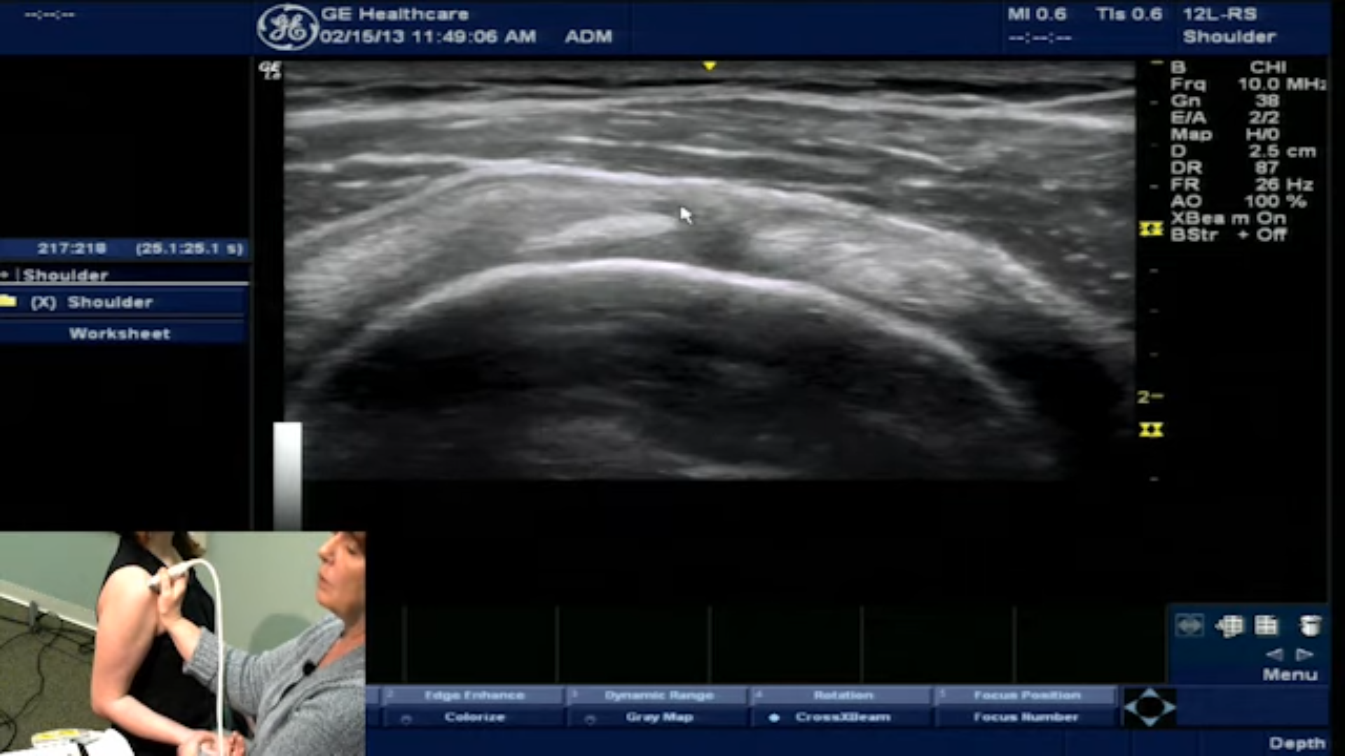
3. Focus on Key Structures:
With the shoulder in this position, you’ll be able to see the main components of the rotator cuff interval. Begin by identifying the subscapularis tendon at the forefront. Right beside it, the biceps tendon becomes more defined. You’ll also spot the supraspinatus tendon and, just above it, the coracohumeral ligament.
Each of these structures has a distinct ultrasound appearance. Becoming familiar with these visuals will not only improve diagnostic precision but also facilitate communication with patients when explaining shoulder anatomy and potential injuries.
Why Position Matters
This positioning technique is a valuable tool for clinicians aiming to visualize the rotator cuff interval clearly. By aligning the tendons and ligaments just right, ultrasound images become more straightforward to interpret, which is especially helpful when diagnosing injuries like rotator cuff tears, tendonitis, or ligament strains. Patients will appreciate the clarity that ultrasound brings to their diagnosis, particularly when they can see and understand the anatomy of their own injury.
The best way to master this technique is to practice! Next time you’re with a patient, take a moment to follow these steps. You’ll soon notice the difference in image clarity and diagnostic potential.
Ready to take your ultrasound skills to the next level? Call the Gulfcoast Ultrasound Institute at 727-363-4500 for all your ultrasound training needs. We’re conveniently located at 111 2nd Ave NE, #800, St. Petersburg, FL 33701. From rotator cuff to advanced cardiac ultrasound, we offer hands-on training and expert instruction tailored to every skill level.