Ultrasonography is an excellent cost-effective modality in imaging of peripheral nerves. High-frequency transducers allow high-resolution imaging at relatively superficial locations. Ultrasound can detect and evaluate inflammatory, traumatic, infective, neoplastic, and compressive pathologies of the peripheral nerves. The normal nerve, in transverse section, reveals small hypoechoic areas separated by hyperechoic septa, displaying a “honeycomb-like” appearance. The hypoechoic areas represent nerve fascicles while the echogenic septa represent the interfascicular perineurium. The longitudinal view also reveals the fascicular appearance described as the “train track” appearance.
System optimization is an essential first step for nerve imaging
- Start with selecting the correct Frequency: High frequency = better resolution. Select the highest frequency that allows penetration through area of interest. Ultrasound evaluation of peripheral nerves should be performed utilizing a high-frequency linear array transducer with a minimum of 7.5 MHz. Higher frequency transducers create better spatial resolution of superficial nerves/structures.
- Make sure your focal zone is set correctly, which is at the depth of the nerves being examined. Some systems are electronically focused, so the focal zone may be automatically adjusted. A single focal zone is used when assessment of a specific level is needed, and multiple focal zones can be used when there is a greater range of depth for the area of concern. Using multiple focal zones will diminish the frame rate and reduce temporal resolution.
- The depth controls the image size, and it is adjusted so that the area of interest takes up most of the field of view.
- Adjusting the Gain/TGC. The ratio of the output to input power is defined as “gain”. The gain amplifies the returning signal to compensate for attenuation. The sonographer adjusts the DGC controls to achieve a uniform brightness throughout the field of view, or in other words, make “like structures look alike.”. The near gain controls the amplification of echoes nearest to the transducer and far gain controls amplification in the far field.
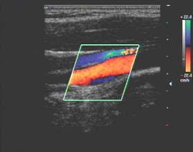
- Color or Power Doppler should be utilized while performing nerve blocks to prevent inadvertent intravascular injection. Doppler is dependent on the angle of insonation so it is important to understand that color Doppler can potentially miss flow in a vessel that is perpendicular to transducer when performing nerve blocks. Unlike color Doppler, power Doppler is NOT direction dependent and is excellent for detecting low or slow blood flow.
Anisotropy in ultrasound is an angle generated artifact created when the angle of incidence is not perpendicular to the structure which results in a distorted and more hypoechoic (darker) appearance of a nerve, tendon Etc. Tilting /angling the transducer to achieve perpendicular incidence to the structure being examined will help to correct this common MSK artifact.
References:
ULTRA P.A.S.S. Sonography Principles and Instrumentation 4th Edition: Green, Lori and Swanson, Mark - Gulfcoast Ultrasound Institute, 2020
Lawande, A. D., Warrier, S. S., & Joshi, M. S. (2014, July). Role of ultrasound in evaluation of peripheral nerves. The Indian journal of radiology & imaging. https://www.ncbi.nlm.nih.gov/pmc/articles/PMC4126140/
GUI has been the leading ultrasound CME provider since 1985. Over 180,000 physicians, sonographers, and other medical providers have attended our programs and utilized our education resources. Post-course surveys reveal a consistent 99% satisfaction rating for meeting or exceeding over-all educational objectives and the learners’ ability to successfully integrate the skills learned into clinical practice.
The Blended Peripheral Nerve Ultrasound Course is designed to provide a comprehensive approach to the use of ultrasound for the assessment and treatment of peripheral nerves. The Blended Education format provides flexibility while providing time and cost savings.
Begin with the self-paced online course: Taught by leading peripheral nerve and musculoskeletal ultrasound experts, the online course incorporates a comprehensive review of the sonographic appearance and recognition of upper and lower limb peripheral nerves, ultrasound guided injection techniques for peripheral nerve injections, regenerative medicine procedures, and the use of diagnostic ultrasound in conjunction with electrophysiologic techniques. The sonographic appearance of muscle for both diagnostic assessment and identification for chemodenervation will also be reviewed.
Then attend Scanning Skills Workshop: Join us in sunny St. Petersburg, FL for a Two (2) Day (11 Hours) scanning skills workshop featuring the industry lowest 3:1 participant to expert faculty ratio with standardized patient models and inanimate phantoms for interventional practice. Because performing ultrasound-guided interventions requires a strong knowledge base pertaining to sonoanatomy, pathology, and scanning and needle-guidance techniques, this course also offers the option to add on a 4-hour interventional cadaver lab with un-embalmed human cadaver specimens to allow the learner to immediately integrate the skills learned into clinical practice.
Visit our website or call us for more information:
111-2nd Avenue NE, Suite 800
St. Petersburg, Florida 33701
727-363-4500