When performing echocardiography, evaluating the tricuspid valve for regurgitation and assessing right ventricular function are critical steps. Today’s hot tip focuses on how to obtain a tricuspid regurgitation (TR) jet and perform a TAPSE (Tricuspid Annular Plane Systolic Excursion) measurement using a modified apical 4 chamber view.
Starting with the Standard Apical 4 Chamber View
- Begin with a basic apical 4 chamber view.
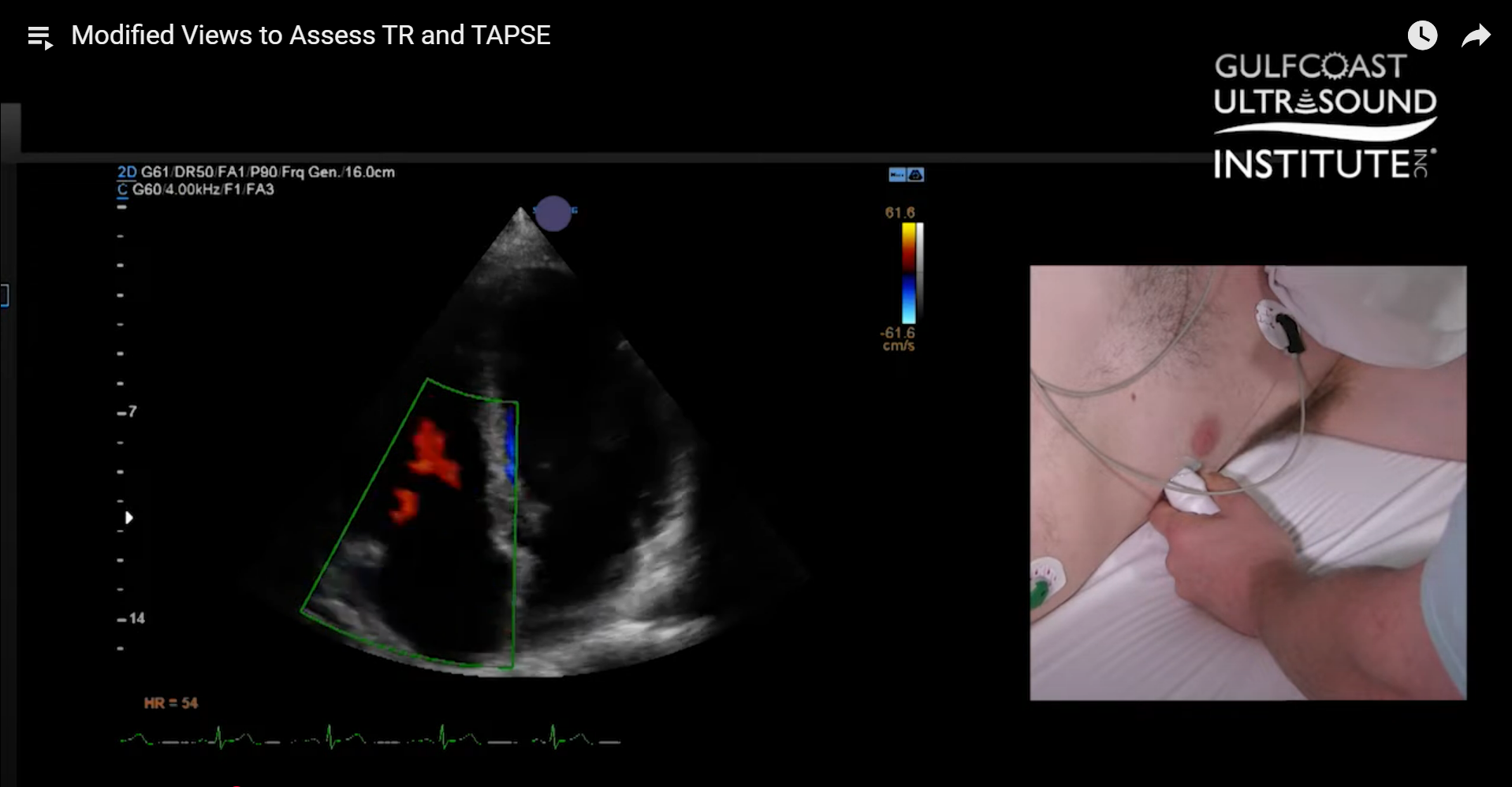
- Apply color Doppler across the tricuspid valve.
- In some cases, the image may appear dark or unclear, making it difficult to visualize regurgitation.
- Use continuous wave (CW) Doppler to evaluate for a TR jet, but remember it may not always be obvious in this standard view.
Why Use a Modified Apical 4 Chamber View?
- Sometimes, sliding slightly off axis provides a clearer window. The key landmark is the relationship between the RV apex and the LV apex:
- If the RV apex sits lower on the screen than the LV apex, you’re on axis for a standard apical 4.
- Sliding off this axis can help bring the tricuspid valve into better view.
- The image often becomes sharper with better probe contact.
- Adding color Doppler reveals hidden TR jets that may not be visible in a standard view.
- Position the CW Doppler cursor directly at the jet’s insertion point to optimize alignment.
- This adjustment allows for a measurable TR jet that might otherwise be missed.
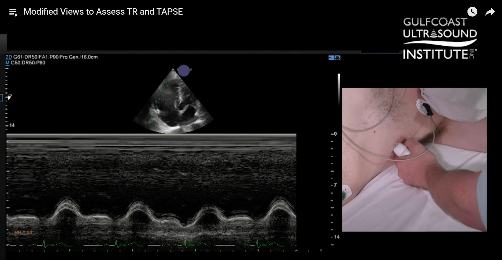
Measuring TAPSE in the Modified View
- TAPSE, or Tricuspid Annular Plane Systolic Excursion, is a standard measurement of RV systolic function.
- From the modified apical 4 chamber view, use M-mode at the tricuspid annulus.
- This alignment places the M-mode cursor more directly with the motion of the RV free wall annulus.
- In a standard 4 chamber view, the RV free wall may obstruct alignment.
- The ASE recommends using this modified approach to achieve a crisp, accurate TAPSE measurement.
Alternative View: Apical 3 Chamber (Reverse 4 Chamber)
- By rotating the probe counterclockwise from the apical position, you can obtain a reverse 4 chamber view.
- Here, the tricuspid valve appears on the right side of the screen and the mitral valve on the left.
- This alternative view can sometimes provide a more parallel alignment for TR jet assessment.
- While not always superior, it is another useful tool in your scanning toolkit.
Key Takeaways
- Always evaluate multiple views of the tricuspid valve when assessing for TR.
- A modified apical 4 chamber view often provides better visualization of TR jets and more reliable TAPSE measurements.
- Don’t forget to try alternative windows such as the reverse apical 4 chamber for additional confirmation.
- Optimizing these techniques ensures accurate calculation of RVSP (Right Ventricular Systolic Pressure) and overall RV function.
Ready to refine your skills and gain confidence in ultrasound-guided injections and procedures like this? Call the Gulfcoast Ultrasound Institute at 727-363-4500 for all of your ultrasound training needs! We’re conveniently located at 111 2nd Ave NE, #800, St. Petersburg, FL 33701.
Whether you’re looking to sharpen your technique or learn new applications, we’re here to help.