Ultrasound-guided procedures have become essential skills for clinicians, enabling precision and enhancing patient outcomes. One critical component of ultrasound needle guidance is the choice between the in-plane and out-of-plane approaches. While many clinicians begin with the out-of-plane approach, which offers its own advantages, the in-plane (or longitudinal) approach provides distinct benefits, especially in situations requiring a higher degree of accuracy. Here, we’ll share a few expert tips on mastering the in-plane approach, helping you overcome common challenges and shorten the learning curve.
Why the In-Plane Approach?
The in-plane approach to ultrasound-guided needle insertion provides a continuous, clear visualization of the needle as it advances toward the target. This technique is particularly beneficial for smaller, more delicate structures like nerves or small vessels, where precision is critical. With the needle fully visible throughout the insertion, clinicians have the advantage of constant, real-time feedback, minimizing the chance of missing the target or causing unintended tissue damage.
The out-of-plane approach remains useful and is commonly employed by beginners. However, developing skills in the in-plane approach opens up new possibilities for procedures such as nerve blocks, vascular access in smaller vessels, and complex injection techniques.
Challenges of the In-Plane Technique
Needle guidance is a fine motor skill that requires precision and stability, much like the intricate movements required for suturing. In the in-plane approach, these skills become even more essential, as the probe is generally held with the non-dominant hand. This can make the learning curve seem steep at first, and it’s not uncommon for new learners to feel frustration as they work to stabilize the probe and control the needle simultaneously.
This technique requires regular practice with gel models or tissue phantoms to refine and reinforce skills. Like mastering a new surgical technique, proficiency in the in-plane approach isn’t achieved overnight or in a single training session. It’s a skill developed over time and with persistence, but the benefits for patient care make it worth the effort.
Key Tips for Successful In-Plane Needle Guidance
- Start with a Clear, Midline View:
When using the in-plane approach, begin by ensuring that you have the target structure centered in your imaging plane. This is particularly important for vascular structures, where precise targeting is essential. To achieve this, use a slight side-to-side movement with the probe. If the structure appears oblique or tapers at the edges of the screen, adjust by twisting or rotating the probe until the vessel is aligned in the longitudinal view, giving you the full length of the target.
- Use Heel-to-Toe Motion for Perpendicular Alignment:
Positioning the vessel perpendicular to the surface of the probe helps create a more defined image of the target structure. Achieve this by gently rocking the probe in a heel-to-toe motion. Once aligned, make sure your probe hand remains motionless to maintain stability and prevent losing sight of the target during needle advancement.
- Stabilize Your Probe Hand for Optimal Control:
After establishing a clear image, maintaining a steady hand is crucial. Stability ensures that the imaging plane remains fixed while the needle advances, allowing for accurate targeting. Good hand stabilization reduces involuntary movements that could disrupt the imaging and lead to misalignment with the target.
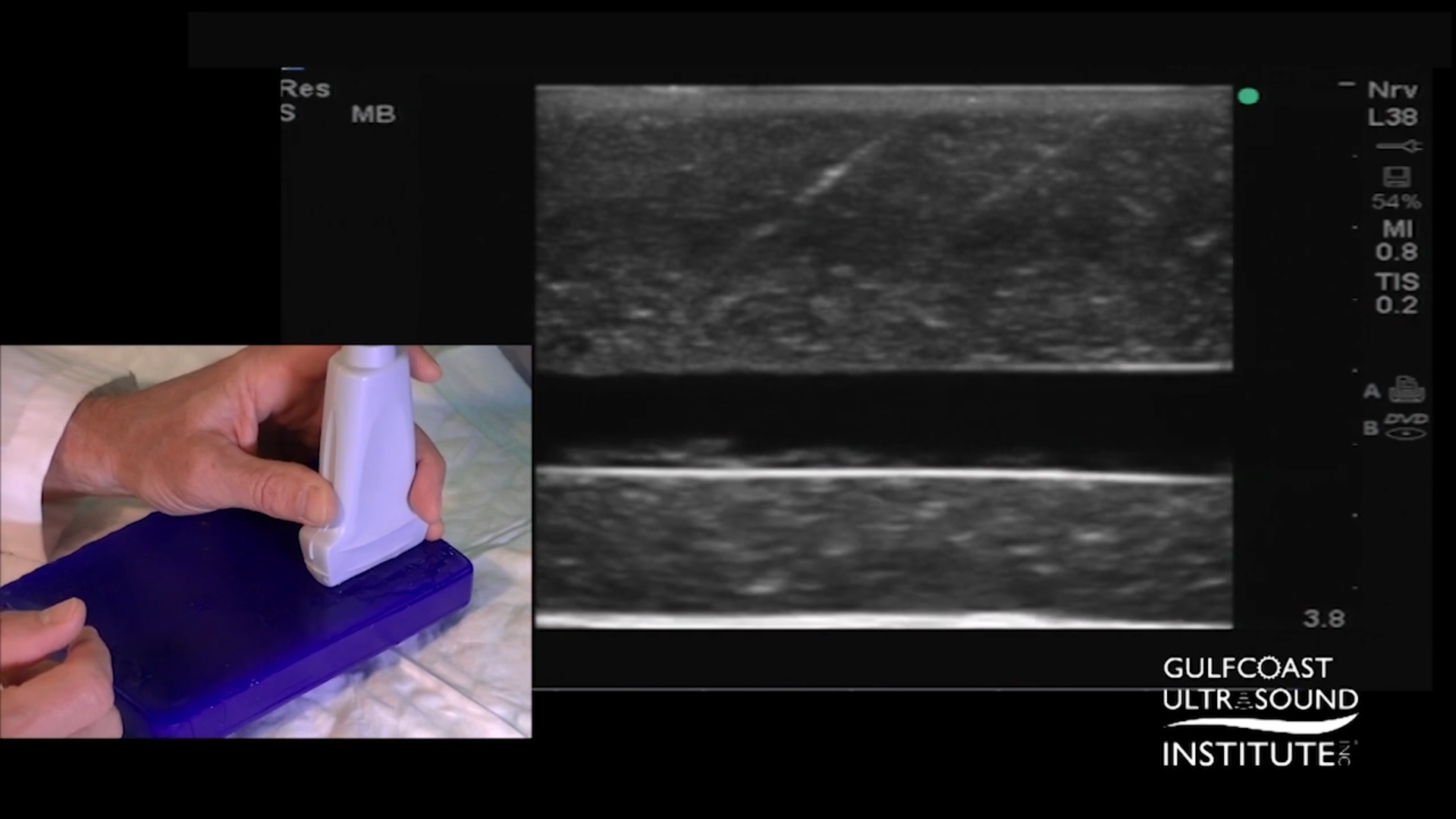
- Approach with a 45-Degree Needle Angle:
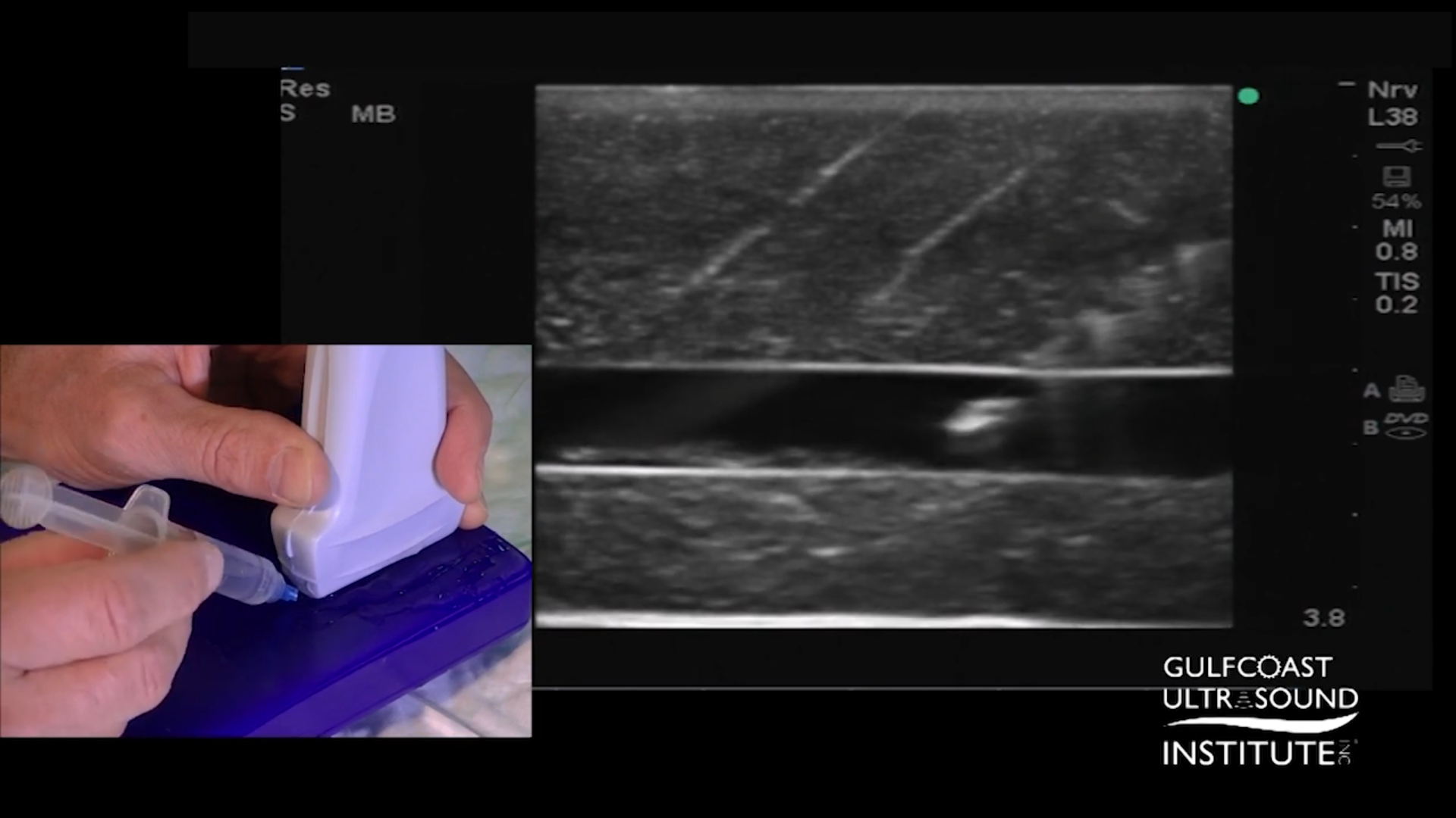
Start advancing the needle from the edge of the probe at a 45-degree angle. At this angle, you’ll see the needle enter from the side of the imaging field, providing a clear visual of the tip. With each advancement, employ a gentle side-to-side fanning motion, which will help you identify the needle tip as it moves through the imaging plane. The goal is to keep the needle tip bright in the image. If the tip remains clearly visible, continue advancing toward the target structure.
- Refine with a Shallower Angle for Precision:
Once in proximity to the target, try lowering the angle to around 15 degrees. This shallower angle offers finer control as you make the final approach. The side-to-side fanning technique remains useful at this stage, helping keep the needle tip centered and bright in the imaging plane. Continue advancing only when the tip is at its brightest, indicating that it’s centered within the plane.
- Final Targeting with Minimal Probe Movement:
For the most accurate insertion, avoid moving the probe once the target is in sight and centered. As you advance the needle, maintain the stabilized position of your probe hand. Any movement could throw off the imaging plane, so consistent hand control is key to a successful procedure.
Practice Makes Perfect
Mastering the in-plane approach for ultrasound needle guidance requires practice, patience, and precision. Regular practice with gel or tissue phantoms can go a long way in refining your skills, helping you build the muscle memory and control needed for complex procedures. Think of it as developing a surgical skill, each repetition builds confidence and expertise, preparing you for real-life applications where precision is paramount.
At the Gulfcoast Ultrasound Institute, we are committed to helping clinicians at all levels advance their ultrasound skills with hands-on, practical training in a supportive environment. Are you ready to take your skills to the next level? Call us today at 727-363-4500 for all your ultrasound training needs! Our center is conveniently located at 111 2nd Ave NE, #800 St. Petersburg, FL 33701. We look forward to guiding you through every step of your learning journey—because mastering these skills today means better patient outcomes tomorrow.