Echocardiography is a valuable tool to provide rapid assessment of cardiac tamponade at the bedside.
Pericardial Effusion
The heart is separated from the surrounding mediastinal structures by a double-walled, fibroserosal sac called the pericardium. The pericardium stops the heart from rotating excessively, prevents friction, and possible kinking of the great vessels. The epicardium or visceral pericardium adheres to the outer heart surface while the parietal pericardium adheres to the diaphragm, inner sternal surface, and adjacent pleural spaces. A space separating these layers contains approximately 5-10 ml of lubricating lymph fluid called the pericardial sac.
Pericardial effusion is defined by an increase in the physiological amount of fluid within the pericardial space. The etiology of pericardial effusion may be due to a variety of medical conditions, inflammation, or cardiac surgery.
2D Echocardiographic Findings of Pericardial Effusion
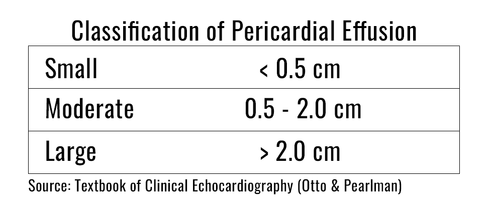
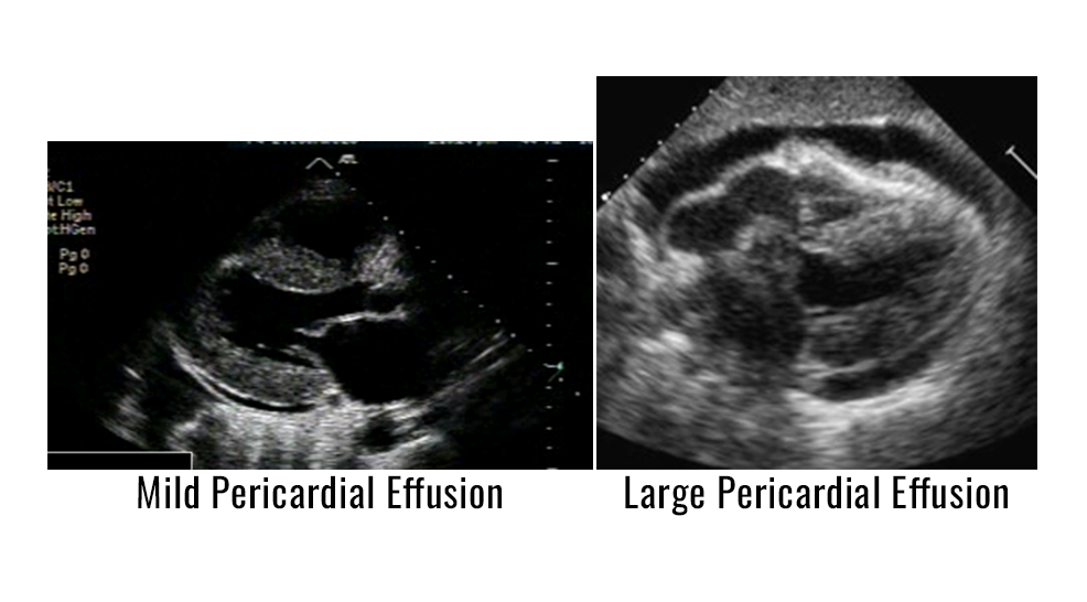
A pericardial effusion appears as an echo- free space of varying size on the 2-D examination. If large enough, the effusion may be seen both posteriorly and anteriorly. In a large pericardial effusion, distant heart sounds producing a dullness in percussion is called Ewart’s sign. In patients with pericardial effusions over a longer length of time, various fibrinous strands may be present and seen in the fluid. Infected or metastatic fluid may appear hazy. When an anterior only echo-free space is noted, a pericardial fat pad may be the cause rather than fluid.
Cardiac Tamponade
Cardiac tamponade is a life-threatening condition that occurs after sudden and/or excessive accumulation of fluid in the pericardial space that results when the pressure in the pericardium is greater than the pressure in the cardiac chambers, resulting in impaired cardiac filling. When tamponade is complete, the diastolic pressures in all of the cardiac chambers are increased and equal to one another. The onset of pericardial tamponade depends on the rate and volume of the accumulation of fluid. Clinical indications of tamponade are tachycardia, hypotension, elevation of jugular venous pressures, and pulsus paradoxus. Pulsus paradoxus is a decline of greater than 10mmHg in systemic pressure with inspiration. Beck’s triad describes three of the clinical indications which includes, increased venous pressure, decreased systolic blood pressure, and muffled heart sounds. Prompt diagnosis is critical to provide the correct patient treatment and management to avoid hypotension and cardiac arrest.
2D Echo Indications of Cardiac Tamponade
- Moderate to large pericardial effusion
- Right atrial systolic collapse that is greater than 1/3 of systole
- Reciprocal changes in volumes of right and left ventricle with respiration
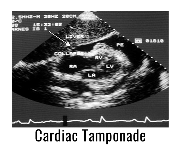
If right atrial systolic collapse is present, it is 94% sensitive and 100% specific for the diagnosis of tamponade. Right ventricular diastolic collapse occurs as a result of intrapericardial pressures that exceed the right ventricular diastolic pressures. This finding can be seen when the right ventricular free wall is normal in thickness and compliance. The finding of right ventricle diastolic collapse is 60-90% sensitive in detecting tamponade, but 85-100% specific as compared to right atrial systolic collapse.
Inferior Vena Cava Plethora
Dilatation of the IVC > 20 mm in an adult patient is an important sign of tamponade. This finding is not specific, but is a very sensitive sign (92%) of cardiac tamponade.
Doppler Evaluation
Doppler assessment of respiratory variations of the transvalvular flows in patients with cardiac tamponade is valuable. Doppler indications of tamponade include:
- Marked decrease (>25%) in mitral valve velocity with inspiration
- Marked decrease (>25%) in aortic velocity with inspiration
- Marked respiratory variation of the pulmonary veins
Summary
Echocardiography is a valuable tool that provides rapid and safe assessment of patients with suspected cardiac tamponade.
References:
Christie Hill Jordan, BS, RDCS, RCS, RCIS, FASE. ULTRAP.A.S.S. Adult Echocardiography Registry Review Workbook, 4th ed. Gulfcoast Ultrasound Institute, Inc.: 2022
Otto.Catherine. Textbook of Clinical Echocardiography, 5th ed. Saunders: 2013
Alejandro Perez-Casares et. al. Echocardiographic Evaluation of Pericardial Effusion and Cardiac Tamponade: Frontiers in Pediatrics. April 2017.
Want to learn more about how to perform and interpret focused or comprehensive echocardiography examinations for improved patient care and safety?
Gulfcoast Ultrasound Institute, Inc. provides multiple CME course options throughout the year. All courses are taught by leading ultrasound experts and offer the lowest participant to instructor ratio (= 3:1) to ensure the maximum amount of skills training and individualized attention. Check out our upcoming 2024 course dates at the link below: